


In ophthalmology clinics of Acıbadem Healthcare Group, each patient undergoes routine eye exam along with contact lens and eyeglass procedures.
Following services are available in examination units; sonography, computer-aided visual field tests, eye angiography and electrophysiological tests. Specialized services include examinations for Retinopathy of Prematurity.
Diagnosis and Treatment Services in Ophthalmology Department
Following diagnosis and treatment services are rendered by Ophthalmology Clinics of Acıbadem Hospitals:
Glaucoma
Diagnosis and treatment services are available for glaucoma in Ophthalmology Clinics of Acıbadem Healthcare Group.
Cataract
Cataract surgery, phacoemulsification procedure and YAG laser, if required, are performed in the unit.
Refractive surgery
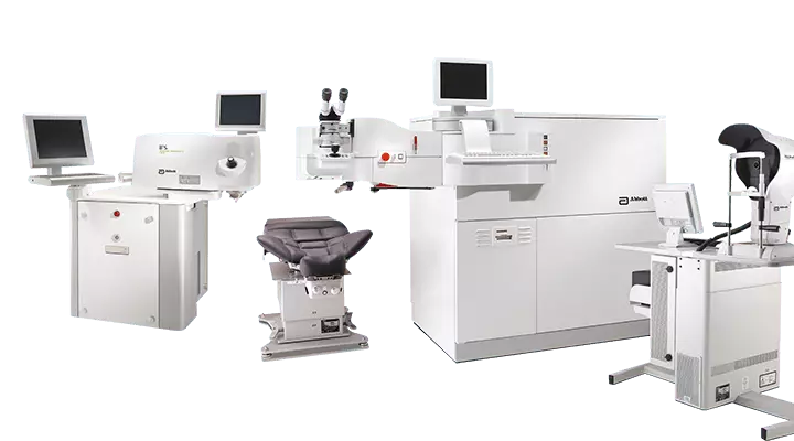
Refractive surgery deals with diagnosis and treatment of diseases that begin in or involve cornea – the outermost layer of the eye. Following devices are used in the unit: topography devices that measure certain parameters precisely; corneal thickness measurement device; specular microscopy; and keratometer that provides data about diameter and refractive power of cornea.
Excimer laser is used to correct refractive errors in Ophthalmology Department. Cornea transplant surgery (keratoplasty) is also performed in the department and patients are recorded on a list; organ transplant surgery is performed, when an appropriate tissue is found through donations or other means.
Oculoplastic Surgery
Cosmetic eyelid surgeries are performed at hospitals and medical centers of Acıbadem Healthcare Group along with drainage surgeries for diseases of eye tear ducts.
Retinal Diseases
Surgical management of retinal detachment and vitrectomy are performed in Ophthalmology departments of Acıbadem Healthcare Group. Argon laser, fundus fluorescein and indocyanine green angiography are available.
Cornea and Contact Lens
Topography devices that measure certain parameters precisely, corneal thickness measurement device, specular microscopy and keratometer that provides data about diameter and refractive power of cornea are available.
Excimer laser is used to correct refractive errors in Ophthalmology Department. Cornea transplant surgery (keratoplasty) is also performed in the department and patients are recorded on a list; organ transplant surgery is performed, when an appropriate tissue is found through donations or other means.
Appropriate contact lenses are selected for each patient and all patients are educated on correct use of lenses in this department.
Strabismus
Strabismus, also called crossed eyes, implies loss of parallel alignment in eyes. There are inward and outward misalignments along with upward and downward ones. Strabismus is broadly classified in two groups; ones secondary to paralysis of eye muscles and others. If strabismus is not secondary to paralysis, sub-categories are congenital strabismus and others.
Lazy eye
If lazy eye is detected, eyesight should be aided by eyeglasses, if required. The department prescribes eye patches and orthoptic therapy along with eyeglasses.
Laser Therapy
Laser therapy options are also available in Ophthalmology departments of Acıbadem Healthcare Group.
LCK
Light Touch CK (LCK) is an FDA-approved modality that is performed at hospitals and medical centers of Acıbadem Healthcare Group. LCK sends radio waves to the marks previously made on eye surface to re-shape cornea. This treatment improves near vision and the treatment should be repeated at 1- to 3-year intervals.
What is cataract? Who are at risk?
Cataract is the loss of transparency in native lens that is located behind iris – the colored part of eye.
Although it is very common at advanced ages;
- Congenital cases are detected in newborn infants,
- It is caused by high blood glucose in diabetics,
- Long-term use of corticosteroids induces the condition or
- It may be secondary to eye traumas.
It is colloquially called “curtain-like vision loss”.
What are symptoms of cataract?- Gradual loss of vision,
- Pale images and colors
- Impaired vision at night
- Difficulty in reading
- Frequent changes in correction power of eye glasses
- Double vision
- Better near vision in certain types (nuclear cataract) Patient specifies that near vision is better. However, it is transient and near vision is also involved, as solidification of lens progresses.
A detailed eye exam is required for diagnosis. Visual acuity is measured in the examination and native lens is examined, after pupil is dilated using an eye drop.
How is it treated?
Treatment of cataract is surgery; medications or eyeglasses do not help the condition. Recently, “Phacoemulsification” technique is used for cataract surgeries.
Types of Cataract Surgeries and Techniques
We use “Phacoemulsification (PHACO)” technique for cataract surgeries. This technique uses ultrasound energy to emulsify the cloudy lens.
Today, “Femtosecond Laser” is used in combination with PHACO technique. This procedure is called “Femto-PHACO”.
How is the surgery done?
Eye is numbed with eye drops and a sedative agent is administered into a vein of arm. Surgery takes approximately 15 to 20 minutes.
First, incisions are made. Next, the cloudy lens is emulsified and aspirated with PHACO and an artificial lens is placed into eye. If Femtosecond laser will be used, side incisions are made and lens is softened with Femtosecond laser.
Is there any risk?
Capsule may perforate, where the cloudy lens is located, and pieces of the cloudy lens may fall into eye. In this case, these pieces are removed in the same session or in a second session.
Postoperative infection of eye (endophthalmia), retinal detachment, corneal edema (swelling of the transparent layer) and Irvin Gass Syndrome (macular edema) are potential complications.
Preoperative
Continue using medications and eye drops that you are already using before the operation. However, you need to inform your ophthalmologist.
Postoperative
Your eye will be examined the next day after surgery. You will be informed by your doctor about the eye drops that you will use for one month. Your eye will be re-assessed 1 week after surgery. An eyeglass examination will be made 1 month after surgery.
If a trifocal lens (a lens that corrects both near and distance visions), you do not need to wear eyeglasses. However, you will need eyeglasses for near vision (reading), if a lens is implanted that corrects distance vision.
Irrespective of the type of lens, it not possible to guarantee 20/20 (perfect) visual acuity. Expectation of a good eyeglass-free vision is 80 to 90 percent.
What should be taken into consideration after cataract surgery?
- The eye drops that are prescribed after surgery should be used regularly.
- Hands should be washed before eye drops are used.
- Eyes should not be rubbed for 1 week after surgery.
- Dirty and foamy water should not contact eyes, while hair is washed.
- Heavy objects should not be lifted in the first week.
- Moreover, you should contact your ophthalmologist immediately in case of severe pain and sleepers. Such pain and sleepers are signs of an infected eye.
Is complete cure possible after cataract surgery?
Certainly. Best visual acuity is expected after surgery, if other parts of eye are healthy. Perfect eyesight can be achieved with or without eyeglasses. If an intraocular lens is implanted, expectation of eyeglass-free vision is 80 to 90 percent.
Laser-aided ocular surgery, type of laser or intraocular lens is decided in the light of a detailed eye exam by your ophthalmologist. This examination also involves detailed evaluation of cornea, where many factors are addressed, such as thickness and morphology of cornea.
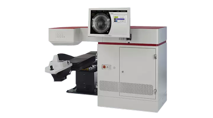
Femto LASIK and Excimer LASER are used for laser-aided ocular surgeries in Ophthalmology Centers of Acıbadem Healthcare Group.
Your surgery is done by our ophthalmologists who are specialized in laser-aided surgery and intraocular lens.
What is Retina?
The outermost part of eye globe is formed by a glass-like transparent layer (cornea) and the colored part of eye (iris). The cavity located behind iris that defines the color of eye, also called “vitreous”, is filled in by a transparent gel.
Retina is a very thin (approximately 0.2 mm) layer that lines the inner wall of globe, just like wall paper.
Why is Retina Important?
Retinal layer consist of millions of nervous cells that receive the light focused by cornea and lens. The light received by these cells is converted into a signal that is transmitted from retina to the brain by optic nerve.
We may speculate that retina, optic nerve and brain are continuum of each other. When retinal nervous cells are completely damaged and cannot sense the light, they cannot regenerate. In other words, irreversible nervous damage causes permanent loss of vision.
Since retinal nervous cells have very high metabolic rate, vascular supply is very crucial; they are fed by both retinal and extra-retinal plexus.
Let’s think retinal nervous cells as pixels of a cell phone. The damaged zone of a mobile cell’s screen looks black and no image can be identified; this example also applies to the damaged zone of retina – the visual field becomes pale, blurred or dark black in the damaged part.
Early detection of a retinal damage and timely treatment of retinal disorders by ophthalmologists who are specialized in retina (eye drops, intraocular drug injections, laser therapies, photodynamic therapy and vitreoretinal surgeries) are very important to prevent an irreversible loss.
What is yellow point? What is its significance?
Yellow point, also called “macula” in medical terminology”, is formed by approximately 1-mm cells at the central zone of retina, where lights are focused on. It is the visual acuity center of eye. These cells sense all details of an image and help us in reading and color vision.
Complete damage of this point results in only five percent of normal eyesight. The central part of visual field cannot be seen. For example, when someone looks at face of a friend, he may see her hair, but cannot recognize the face.
What are Retinal Diseases?
Retinal nervous cells or their feeder vessels may impair. Causes of these disorders may change by age of the person. Other systemic diseases may also affect retina and feeder plexus.
Common Retinal Diseases:
- Age-related degeneration, bleeding and edema in the yellow point (age-related macular degeneration)
- Retinal degeneration that is predisposed to tear, retinal tear, and retinal detachment
- Retinal hemorrhages, edema and retraction in yellow point, vitreous hemorrhages and retinal detachment in diabetic patients, especially with poorly regulated blood glucose,
- Retinal hemorrhages, obstruction of feeder arteries or veins that carry oxygen-poor blood, bleeding secondary to systemic hypertension,
- Diseases secondary to disorders at the junction of the yellow point and the gel (vitreous), a hole in the yellow point, membrane formation, retraction and edema of the yellow point,
- Accumulation of fluid in the yellow point due to various causes,
- Tear of retina and other layers secondary to blunt or sharp traumas, retinal detachment, vitreous hemorrhage, foreign body in eye, infection of eye, injury of lens and detachment of lens into vitreous.
- Bleeding in preterm babies secondary to immaturity of retinal blood vessels, retinal detachments that may progress even to loss of the eye,
- Genetic diseases that are associated with gradual damage to retinal cells over years; retinitis pigmentosa is the most common one of these conditions,
- Benign and malignant tumors of retina or retinal blood plexus and metastasis of cancers that begin in other body parts, albeit more rarely.
What are Symptoms of Retinal Diseases?
The person should test visual acuity of near and distance vision separately for each eye. Binocular vision may cause late recognition of the poor vision confined to one eye.
This simple self-examination is made as follows; select an object or a distance and close one eye in each evaluation to determine visual acuity of both eyes separately.
If one of the following symptoms is recognized in near or distance vision, a retinal disease should be suspected and medical attention should be sought.
- Blurred vision,
- Blurred/dark central zone of visual field,
- Blurred, cloudy or pale vision in any part of visual field while looking at somewhere,
- Inability to distinguish color tones or difference between two eyes,
- Straight lines, rows (while reading) and window and door borders look wavy and distorted,
- Seeing flashing lights in the dark,
- Floating spots, threads or other shapes that suddenly appear in eyesight.
Amsler grid is a test that consists of a chart with numerous small squares that are identical in size; this chart allows self-examination by closing one eye in each turn.
For the test, the person wears farsightedness or reading glasses, if any, or looks at the central point that he sees best by his age (no eyeglass can be worn by myopic patients) and finally, the person checks whether all squares are properly seen and whether there are dark areas in eyesight. This test is specific to yellow point diseases and it does not evaluate other retinal areas.
You should immediately visit an ophthalmologist who is specialized in retina, if you detect any abnormality in Amsler test or if you have any one of the above written complaints in self-examination of near and distance vision separately for each eye.
How are Retinal Diseases Treated?
Since there are numerous disorders of retina, treatment may vary by the condition and the time since onset of complaints.
Basic options:
Eye drop,
Intraocular drug administration:We use this treatment option very commonly. It is especially employed for age-related macular degeneration and edema of the yellow point due to any reason. Since drug is administered into vitreous, it affects retina earlier and more efficiently. Intraocular therapeutic effects of these drugs continue around one month. Repeated treatments may be required.
Argon laser photocoagulation:
It is used for many retinal diseases, such as diabetes-related retinal disorders and vascular occlusions.
How is it applied?
The patient is seated on a chair that is similar to the one used for slit-lamp examination. Eye is numbed with an eye drop. The laser light is focused on retina using a lens and the patient only hears a sound. No pain or ache is felt. The procedure usually lasts for 10 to 20 minutes and the patient can be discharged without an eye patch. Routine activities of daily life can be resumed on that day. Only blurred vision persists for several hours, as pupil is dilated before the procedure.
Photodynamic Therapy:
It is similar to laser therapy. Today, it is not very commonly used, as intraocular drug administration is more effective. For necessary conditions, an intravenous drug is administered and cold laser is applied. It is expected to affect the underlying vascular tissue with no damage to retina. Patients should protect eyes and other exposed areas of body from light for 48 hours after the procedure. The patient is prescribed a protective eyeglass.
Retinal Surgeries or Vitreoretinal Surgery (Vitrectomy with microsurgical technique):Retinal surgeries are performed by ophthalmologists who are educated on these surgical techniques. An ophthalmologist should specialize on retinal surgery following standard ophthalmology residency.
The patient is informed by retina ophthalmologist about the surgery technique and timing of the operation.
Which Diseases Are Managed with Retinal Surgeries?
They may be required for numerous retinal diseases. Primary examples include retinal detachment, intraocular hemorrhages, hole of the yellow point, membrane formation, retraction and resultant edema, retinal tractions secondary to uncontrolled diabetes and eye traumas.
How are retinal surgeries performed?
They are performed in operating theaters under general or local anesthesia. These techniques are as follows:
a. Vitreoretinal Surgery (Vitrectomy with microsurgical technique):It is the most commonly used technique. Vitrectomy is performed with closed technique, similar to laparoscopic abdominal surgeries. Cannula are inserted into eye through three points in conjunctiva – the white part of eye – and the problems that involve vitreous and retina are eliminated, such as correction of retinal detachment and peeling microscopic membranes that form on retina, or argon laser is applied, if necessary. The cannula used for vitrectomy measure approximately 0.5 mm in thickness.
A fluid, air or gas or silicone oil is injected into and left in eye, when this surgery is completed. The patient is preoperatively informed in detail. If the patient has cataract that may potentially influence the success of vitrectomy in technical terms, a cataract surgery can also be performed in the same session.
b.Cerclage, Buckling Technique:It is an extraocular surgery technique. Some conditions may require combination of this surgery with vitrectomy.
Thin silicone strips (usually 3 to 5 mm thick) are advanced beneath conjunctiva – the white part of eye – and placed under ocular muscles.
Re-attachment of retina is expected through external compression of eye. It is combined with either Argon laser or cryotherapy (permanent attachment is created through freezing the damaged part).
c.Pneumatic Retinopexy (intraocular gas injection):We do not use this technique very commonly. Because an appropriate case is rarely admitted.
For eyes with retinal detachment that is associated with tear, gas is injected into eye in the early stage to allow correction of detachment while the patient rests in an appropriate position. It is followed by argon laser therapy.
Intraocular gas injection is also used for macular hemorrhages to remove the accumulated blood.
How About Recovery after Retinal Surgery?
The patient is preoperatively informed about postoperative expectations in detail.
- If gas or silicone oil is injected into eye, the patient should sleep/sit in prone position for a period that is determined by the surgeon (may vary from 1 day to 2 weeks).
- As is the case with postoperative period of cataract surgery, eye drops are used for one month in gradually reduced doses.
- The first week is especially important regarding prevention of infection. It is very important to avoid too crowded and dusty or dirty places and not to touch or rub eyes.
- It is optimal to have sick leave and rest for one week after surgery. This period may vary depending on severity of the condition.
- If air or gas is injected into eye, flights and travel to high altitudes are strictly forbidden. One must necessarily consult the surgeon in advance.
- It is also reasonable to avoid heavy sports, weight lifting, strenuous works and spraining and straining, such as cough, for one month after surgery.
Eyesight may vary depending on the underlying cause of disease. Your doctor will inform you in detail about this issue.
Hospitals
-
 Adana Hospital
 -
 Altunizade Hospital
 -
 Ankara Hospital
 -
 Atakent Hospital
 -
 Ataşehir Hospital
 -
 Bağdat Outpatient Clinic
 -
 Bahçeşehir Outpatient Clinic
 -
 Bakırköy Hospital
 -
 Beylikdüzü Surgical Outpatient Clinic
 -
 Bodrum Hospital
 -
 Bodrum Outpatient Clinic
 -
 Bursa Hospital
 -
 Dr. Şinasi Can (Kadıköy) Hospital
 -
 Eskişehir Hospital
 -
 Fulya Hospital
 -
 Göktürk Outpatient Clinic
 -
 International Hospital
 -
 İzmir Kent Hospital
 -
 Kayseri Hospital
 -
 Kent Bayraklı Medical Center
 -
 Kozyatağı Hospital
 -
 Maslak Hospital
 -
 Taksim Hospital
 -
 Zekeriyaköy Outpatient Clinic
Doctors
-
 Prof. ALİ RIZA CENK ÇELEBİ, M.D.
-
 Prof. ALTAN GÖKTAŞ, M.D.
-
 Prof. AYŞE ÖNER, M.D.
-
 Prof. BANU COŞAR, M.D.
-
 Prof. BERNA ÖZKAN, M.D.
-
 Prof. DİLAVER ERŞANLI, M.D.
-
 Prof. DİLEK GÜVEN, M.D.
-
 Prof. G. ERTUĞRUL MİRZA, M.D.
-
 Prof. HALUK ESGİN, M.D.
-
 Prof. İBRAHİM BÜLENT BUTTANRI, M.D.
-
 Prof. KEMALETTİN KAZIM DEVRANOĞLU, M.D.
-
 Prof. MEHDİ S.ÖĞÜT, M.D.
-
 Prof. MUHSİN ERASLAN, M.D.
-
 Prof. MÜSLİME AKBABA, M.D.
-
 Prof. NAZAN BENGÜDENİZ ERDA, M.D.
-
 Prof. ÖMER CAN ÜSTÜNDAĞ, M.D.
-
 Prof. ÖZGÜL ALTINTAŞ, M.D.
-
 Prof. SARPER KARAKÜÇÜK, M.D.
-
 Prof. SELÇUK SIZMAZ, M.D.
-
 Prof. SOLMAZ BALCI AKAR, M.D.
-
 Prof. TUNÇ OVALI, M.D.
-
 Prof. U.EMRAH ALTIPARMAK, M.D.
-
 Assoc. Prof. AYŞE EBRU BAHADIR, M.D.
-
 Assoc. Prof. NESLİHAN SİNİM KAHRAMAN, M.D.
-
 Assoc. Prof. ÖZGE BEGÜM COMBA, M.D.
-
 Assist. Prof. FATİH ATMACA, M.D.
-
 Assist. Prof. İBRAHİM ŞAHBAZ, M.D.
-
 AYTEK COŞAR, M.D.
-
 BÜLENT RAŞİT TUNCEL, M.D.
-
 DENİZ MARANGOZ, M.D.
-
 DİLEK TOMATIR, M.D.
-
 DİLEK ABUL, M.D.
-
 EMEL ÇOLAKOĞLU, M.D.
-
 EMRE ÇAKIR, M.D.
-
 EMRE SÜBAY, M.D.
-
 ERBİL ULUS DUMAN, M.D.
-
 ERCAN PAŞAOĞLU, M.D.
-
 EVREN BACA, M.D.
-
 GÜLHAN SARIAYDIN, M.D.
-
 HÜLYA DEVECİ, M.D.
-
 MEHMET KANTARCI, M.D.
-
 MELİKE LİDER GEZER, M.D.
-
 MUSTAFA DEMİR, M.D.
-
 MÜRÜVVET AYTEN TÜZÜNALP, M.D.
-
 NEZİH ÖZDEMİR, M.D.
-
 NİLAY AKAGÜN, M.D.
-
 ÖKKEŞ BAZ, M.D.
-
 ÖMER FEYYAZ KADIOĞLU, M.D.
-
 SAFİYE KÜÇÜKGÜL, M.D.
-
 SEVDA ARIK TEKİN, M.D.
-
 SUZAN KARAMETE, M.D.
-
 ŞAFAK ÇAKMAKÇI, M.D.
-
 TOLGA YÜKSEL, M.D.
-
 UĞUR YILMAZ MUMCU, M.D.
-
 YELDA ERTÜRK, M.D.
-
 YEŞİM TASALI, M.D.
-
 YUSUF İZZET GÜNGÖRMÜŞ, M.D.
-
 ZEKİ ERMİŞ, M.D.