PET-CT is employed especially in oncology (cancer) to identify the tumor, determine the spread of tumor, plan radiotherapy, evaluate chemo- or radio-sensitivity and decide whether the mass lesion is benign or malignant.PET-CT utilizes the increased glucose metabolism, which is a characteristic of cancer cells. A glucose molecule labeled with radioactive substance is administered through intravenous route before the scan and this molecule accumulates in cells that form the tumor.
Since whole body is scanned, it can determine certain conditions like spread of cancer to other parts of body (metastasis).
Moreover, the test is utilized for diagnosis, staging and re-staging of metastases with unknown origin.
PET-CT is used mainly for following cancers:
- Lung cancer
- Breast cancer
- Lymphoma
- Gynecologic cancers
- Colorectal cancers
The test can also be used for imaging other less common cancer types.
PET-CT also plays role in neurologic cases for determination of epilepsy focus, early diagnosis of Alzheimer's disease and investigation of viable tissue in heart after a heart attack.
PET-CT for Early Diagnosis
Many diseases can be diagnosed early with PET-CT. While biopsy was the only option to determine whether a nodular formation is cancer or not before PET-CT was introduced to clinical use, PET/CT can accurately clarify if these lesions are cancer.
As the data on body images is obtained using computer systems, even the tumors measuring 5 mm in diameter can be visualized with PET-CT.
PET-CT at Metastasis Stage
It is also used to detect in many cancers whether the cancer spreads to nearby tissues or lymph nodes (metastases).
Since the entire body is visualized at the same time, it is possible to investigate whether the disease spread to another organ. For example, it is possible to reveal out whether there are metastases of a lung tumor in bone, other visceral organs, adrenal glands or lymph nodes, or in other words, stage of the disease is determined.
PET-CT after Therapy
PET-CT has an important role in imaging phase, as it provides detailed information about the tumor. PET can render images of metabolism in tissues and therefore, after presence of a lesion is anatomically verified, it can be understood whether it is active regarding malignancy.
For example, lymph nodes enlarge in lymphoma and they are reduced with treatment, but they may not completely disappear.
Enlargement of lymph nodes may persist to an extent in most cases after the treatment. It is known that PET-CT is the most important diagnostic tool to understand if there is still an active disease in them or they have completely disappeared. Therefore, PET-CT not only locates the lymph nodes, but it also helps determine whether there is still an active disease or there is complete response to therapy.
Due to these reasons, PET-CT helps the physicians regarding early diagnosis of cancer and saves time which is necessary and critical for treatment.
Clinical Indications of PET-CT
Lung
PET-CT has a critical role in investigation of lung cancer. A cancerous tissue is considered by 96 percent, if this imaging modality demonstrates increased F-18 Fluorodeoxyglucose uptake in nodules that are located in lungs.
Evaluation of tumor, regional lymph nodes and presence of metastasis are of great importance in staging lung cancers before treatment. These data are acknowledged as a guide to plan the treatment.
It is possible determine recurrence of surgically removed lung cancers precisely with PET-CT.
Identification of distant metastasis is among other clinical indications of whole body PET-CT along with determination of relapse.
Distant metastases can be detected at any stage of treatment and they require modification of the treatment approach.
Lymphomas
Practices with PET-CT gain ever-increasing importance for Hodgkin's and non-Hodgkin's lymphoma. Sensitivity and specificity is 90-100 percent for diagnosis of lymphomas in comparative studies conducted with anatomic imaging methods.
As the increased glucose metabolism in anatomically visualized lymph nodes indicates active lymphoma, ability to scan whole body with PET-CT provides additional advantages. Moreover, the lesions can be visualized with PET-CT in case of unusually located lymphoma such as primary small intestinal lymphoma.
Staging plays a critical role in selection of treatment for lymphomas. Uptake in thoracic or abdominal organs and pelvic lymph nodes has vital importance in determination of treatment method.
Thyroid cancers
Although PET-CT is not one of the conventional diagnostic tools in thyroid cancers, it can be preferred by physicians in case of suspicious lesions. PET-CT can be scanned in case of problems in sufficient tissue sampling for histological examination and especially determining benign and malignant follicular nodules in fine needle aspiration biopsy.
PET-CT is very important in detection of recurrence or metastatic spread especially for patients who have thyroid cancer with negative I-131 whole body scan.
For the patients with no I-131 uptake but high thyroglobulin level, it is reported that PET-CT has high sensitivity in detection of lesions.
Breast Cancers
While physical examination and mammography are most commonly used for diagnosis of breast cancer, certain problems can be faced due to some reasons such as dense breast, mammoplasty and prior needle biopsies.
PET-CT can be used for diagnosis in case of small lesions, indefinite mammography findings and fibrocystic diseases in addition to above mentioned problems.
While breast cancer is still the most common cancer in women around the world, the condition of axillary lymph nodes is the most important predictive factor. Detecting metastatic axillary lymph nodes with PET-CT is of great importance for treatment.
Pancreatic Cancers
Primarily manifested by abdominal pain and weight loss, pancreatic cancers are usually diagnosed late. For diagnosis of pancreatic cancer,
Blood tests, MRI and computerized tomography are used. Tumor markers such as CA 19 9 and CEA are usually high in blood tests of patients. PET-CT is scanned to detect malignant tumors.
Sensitivity of PET-CT to differentiate pancreatic adenocarcinomas from benign pancreatitis (inflammation of pancreas) that has mass effect is 94 percent. Moreover, PET-CT provides critical data in staging of pancreatic cancers and investigation of metastases.
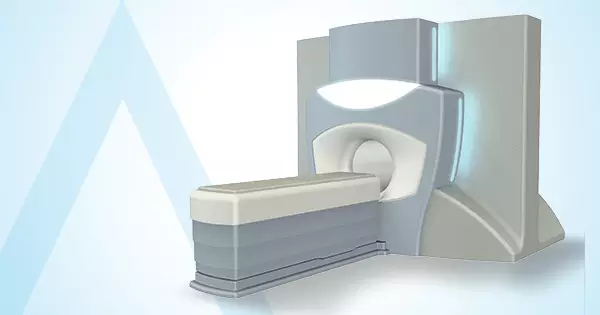
For treatment of pancreatic cancer, MR-LINAC can irradiate and destroy the tumors, which are located in organs that move during treatment, with pinpoint accuracy. Tumors can be monitored closely and destroyed with pointed radiation therapy during surgery as radiation beams are delivered with MRI guidance.
Successful outcomes are obtained in pancreatic cancer in treatments that follow MR-LINAC therapy. It is reported that the success rate has increased from 10 percent to 90 percent, after MR LINAC was introduced to therapeutic use.
Colon cancers
Sensitivity of PET-CT is significantly high for diagnosis of colorectal cancers, which account for approximately 13 percent of all malignancies (malignant tumors). However, PET is not commonly used in diagnosis of primary colon cancers or initial staging in routine practice, unless metastases in or out of liver are not investigated.
Colorectal cancers can be diagnosed with 90 percent sensitivity using affordable and common methods such as colonoscopy and double contrast barium enema. It is known that PET-CT is beneficial for detecting colonic adenomas regarding diagnosis.
Colorectal cancers are one of the malignancies with highest recurrence rates. Frequently, recurrence is found in 30-40 percent of cases within two years after treatment. The condition is only seen as liver metastasis in some patients. However, number of relapsed foci and extrahepatic spread are among the important factors for treatment. PET-CT gains importance as MRI in addition to CT cannot provide sufficient benefits in differentiation of postoperative scar tissue and recurrence.
In the light of all these information, PET-CT becomes a useful choice if there is suspicion of recurrent colon cancer, tumor markers are high although there is no known focus, and recurrence cannot be verified with conventional imaging methods and for preoperative staging before surgical removal of recurrent tumor.
Melanoma (Skin Cancer)
Melanoma is the most aggressive type of skin cancer. It may metastasize to every organ, although regional lymph nodes are involved earlier.
Although treatment includes surgical removal, lymph node dissection is preferred for diseases with metastatic spread due to complication rate of 5 to 40 percent.
PET-CT can determine condition of regional lymph nodes and whether there is metastasis in other organs with high accuracy rate.
Brain Tumors
PET-CT is also used for brain tumors. Certain changes can be seen in brain tissues of patients, who receive radiotherapy, due to radiation necrosis after a certain period especially following the surgical management of brain tumors. PET-CT is scanned to understand if these changes are secondary to tissue necrosis due to radiation or recurrence of tumor.
Other Fields of Use
In addition to these common clinical indications, PET-CT is scanned for following diseases:
- Head and neck tumors,
- Mesothelioma,
- Mediastinal lesions,
- Esophageal cancer,
- Stomach cancer,
- Small intestinal tumors,
- Gastrointestinal stromal tumors,
- Hepatobiliary tumors,
- Gynecologic malignancies,
- Genito-urinary system tumors,
- Bone tumors,
- Soft tissue sarcomas,
- Neuroblastoma and metastatic tumors with unknown primary.
Critical information regarding diagnosis, staging and treatment response is gained for these diseases.
Moreover, metastases in almost all organs can be detected with PET-CT, more commonly in lymph nodes, lung, liver and bone.


